
DPM, MPH
PRESENT Editor,
Diabetic Limb Salvage
Diabetic Foot Ulcers: Don’t Forget the Basics!
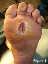
The most characteristic lesion associated with the diabetic foot is the foot ulcer (Figure 1). Diabetic foot ulcers (DFU) are certainly nothing new – they are vividly described in very old medical literature exactly as we would describe them today. Adjectives such as painless, deep crater-like center, hypertrophic callus on the borders, with varying degrees of granulation and necrotic tissue come to mind from the description by Mott published in 1818.1 With the increasing number of persons with both type 1 and type 2 diabetes mellitus (DM) around the globe, the numbers of associated foot ulcers will also show a commensurate increasing frequency. Indeed, every region in the world already seems to be burdened with this chronic complication of diabetes. Most importantly, we must recognize the close association between foot ulcers, infections, and amputations in this at-risk population. Pecoraro published his classic study of component causes leading to diabetes-related amputations in 1990.2 The three most common component causes found in the causal pathway to amputation included neuropathy, ulceration, and wound healing failure. In fact, 85% of the amputations studied had a foot ulcer in the pathway leading to the amputation. This is not to infer that 85% of DFUs lead to amputation. However, several studies have indicated that 15 to 20 percent of ulcers go down the pathway to amputation due to subsequent infection and/or ischemia.3,4
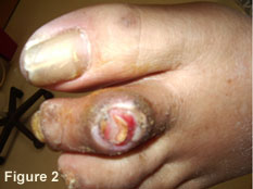
The causal pathway to diabetic foot ulceration was similarly studied and published in 1999.5 Among neuropathy, ischemia, infection, callus, deformity, and trauma, the most frequent constellation of component causes found in this study were neuropathy, deformity, and trauma. This is not surprising, since neuropathy seems to be the critical component underlying most diabetes related lower extremity complications. A very common clinical scenario that exemplifies this pathway would involve that typical long- standing diabetic patient with loss of protective sensation (LOPS) who has a simple hammer toe deformity. After wearing a new pair of shoes for several hours, a blister had developed on the toe and was unrecognized until that night when the patient removed the shoes and noticed the bloody stain on his socks. This is the good scenario. All too often, people with LOPS don’t recognize the wound until infection has set in or it starts to smell. (Figure 2) In these late cases, hospitalization is usually required, as is surgical drainage or debridement.
Recognition of associated risk factors for DFU helps us manage the lesions more effectively, as well as to assist in the prevention of new or recurrent lesions. While there have been numerous other studies investigating putative risk factors for DFU (i.e. HgbA1c, smoking, prior amputation, PAD, education, footwear, etc.), the basic tenets of treatment for any DFU remain the same.6 While simplification is not always good in the management of patients with potentially limb threatening ulcers, condensing the basic components of care facilitates their systematic implementation. Of course, a thorough systematic history and examination is a necessary precursor to treatment. For instance, if one does not systematically consider the influence of underlying ischemia in a chronic non-healing wound, he/she might not do a thorough diagnostic evaluation for vascular disease. The same is true for occult osteomyelitis underlying a chronic wound of long duration. Much has recently been written about the efficacy of advanced therapies in the management of recalcitrant ulcers.6,7,8,9,10 Nonetheless, advanced therapies must be considered as adjunctive to standard, good wound care. Would anyone consider applying a tissue substitute product to an infected, ischemic wound? Of course not, because the treatment would be doomed to failure unless the primary components of infection and ischemia had been first resolved.
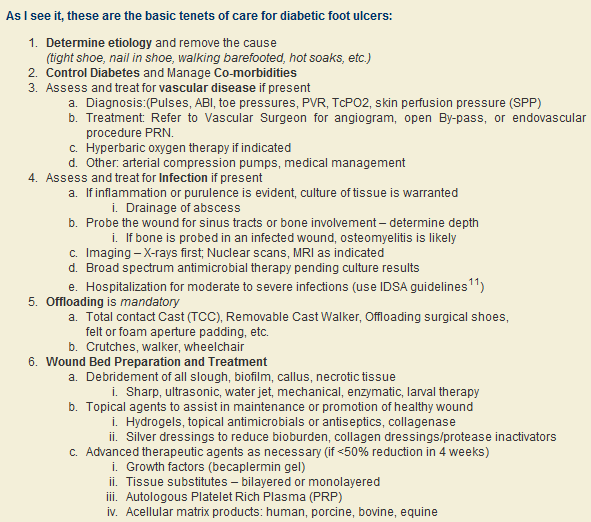
By no means is this listing of diagnostic and treatment parameters meant to be exhaustive. To the contrary, it is meant to be rather straightforward and logical so that it can be easily remembered. After all, you won’t always have a wall chart or pocket guide in front of you as you are assessing a patient and determining the extent of pathology and necessary treatment for a diabetic foot ulcer. I can honestly say that for every such patient that I treat, these same six basic tenets come to my mind. I go through them systematically, so that I can be sure to evaluate the important parameters and provide the necessary treatment regimen. A thorough, systematic evaluation will ascertain the important underlying pathology and will therefore facilitate appropriate treatment. It’s really that simple (if managing a diabetic foot lesion can really be considered simple)! Treating the majority of these wounds does not have to be a daunting or complex task. Sometimes, the most important aspect is to make the proper assessment and then, as circumstances warrant, make the appropriate referrals. It is well established that a multidisciplinary team approach will lead to the best outcomes for diabetic foot ulcers.12
Although classification of the DFU has not been mentioned in this FootNote, a proper evaluation and subsequent treatment will still most often lead to a successful outcome. Nonetheless, there are several wound classification schemes in common usage around the world that can not only classify a wound, but also aid in appropriate communication, treatment, and prediction of outcomes. That will be a topic of a future FootNote.
References are provided below that can expand upon many of the points made above. We welcome your opinions, concerns, and suggestions.
Best regards,
Robert Frykberg, DPM, MPH
PRESENT Editor,
Diabetic Limb Salvage
REFERENCES
|