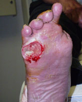
Classifying Diabetic Foot Ulcers In the December 2010 FootNotes, we discussed the basics for managing diabetic foot ulcers (DFU). I’ve always believed that with a thorough, systematic approach to the examination of DFUs, one could appropriately institute treatment by addressing the basics — as well as those parameters found to be abnormal. For instance, if one does not look for ischemia in the neuropathic patient, the clinician can easily miss this important parameter that can impair wound healing. The neuropathic diabetic patient will often not have any symptoms of claudication or rest pain that we rely upon to trigger the suspicion for peripheral arterial disease (PAD). Similarly, infection can predict failure to heal. While active infection is fairly easy to diagnose, occult or smoldering underlying osteomyelitis often eludes diagnosis because there are often no commensurate laboratory or radiologic signs to herald its presence. Numerous studies have indicated that larger ulcers and those with a long duration are more likely to have concurrent underlying osteomyelitis. Therefore, I think that it is critical to make the diagnosis of these two common complicating factors when assessing DFUs on their initial presentation. Classification – Why Do We Need It? Although time and space preclude a detailed discussion, there are several well accepted classification systems for DFUs. The purpose of such classification is not only to communicate severity or status of a wound, but also to facilitate treatment. For instance, if one diagnoses bone involvement indicative of osteomyelitis then that diagnosis portends a certain level of risk for failure or even amputation. This, of course, relies upon looking for this complication in an otherwise non acutely infected foot ulcer. Furthermore, once the ulcer is classified as having bone infection, treatment must then follow. Along the same lines, once underlying PAD is diagnosed or suspected in a DFU patient, it is then incumbent upon the clinician to confirm the diagnosis and refer the patient for possible revascularization. Again, one must investigate carefully and look for this significant complication when assessing the patient presenting with a new or chronic foot ulcer. It really does not have to be complicated — with a thorough examination the appropriate treatment naturally follows. This is the value of classifying wounds. The classification systems require that the clinician evaluates these parameters in order to place the wound in various categories or grades. Progressively increasing grades of severity (or depth for instance) portend the need for more aggressive treatments as much as they portend risk for wound healing failure and amputation. The Classification Systems – What is Most Helpful? One of the older and perhaps more well known classifying schemes is that proposed by Wagner (and Meggit) in the 1970s. The Wagner system has six grades, from 0 to 5, representing the foot at risk for ulcer (0) to an entirely gangrenous foot (Grade 5). The extent of tissue loss or depth of penetration is well described, but the important concurrent parameters for infection and ischemia are missing from this scheme (not that I agree that gangrene should be classified as an ulcer). Nonetheless, partial foot gangrene (grade 4) requires at least a partial foot amputation and a Grade 3 lesion that penetrates to bone or joint will require further investigation and management for likely osteomyelitis (Figure 1).
The shortcomings of this system lead to the development of theUniversity of Texas system in the mid 1990s. In this more thorough, but complicated system, infection and ischemia play important parts in the classification scheme that is really based on the Wagner system. Instead of six categories, or cells to remember, the UT System has 16 cells made of a grid (4 x4 table) consisting of 4 grades/depths and 4 stages (neuropathic (A), infected (B), ischemic (C), and (D) infected with ischemia). While this can become complicated to the unacquainted clinician, it has been validated as being predictive for amputation with increasing grades and stages (3D implying a wound penetrating to bone with both infection and ischemia). In addition to the UT System, the PEDIS system was developed in the last decade to be a more specific scheme to assist in the classification of DFUs for research purposes. Perfusion,Extent/size, Depth/tissue loss, Infection and Sensation are the components of interest in the PEDIS system. Again, the number of cells and complexity of the latter system make it difficult to use extemporaneously in clinical practice. On the other extreme, an overly simplistic approach such as mild, moderate, and severe is just too broad and nonspecific as to be of any great value. With all the aforementioned being recognized, I tend to simplify things as much as possible (it’s easier to remember and I do not need a wall chart to classify the wounds in front of me). All of the parameters already mentioned are incorporated into my evaluation and subsequent treatment plan. To me, however, classifying wounds as Neuropathic, Ischemic, or Neuroischemic facilitates treatment very nicely, especially with modifiers indicating infection and depth. In the most basic of terms, an infected 3 cm neuroischemic ulcer with exposed bone tells me all that I need to know in order to proceed with treatment. Furthermore, I know that there will be a high likelihood for osteomyelitis that will need to be treated once the patient has been referred out for revascularization. Granted, this is a simplistic approach, but one based on fully evaluating the parameters required for the more thorough classification systems. It works for me, although it may not be precise enough for interprofessional communication, validation, or for research purposes. The point is to thoroughly and systematically assess DFUs and upon finding or suspecting important concurrent pathology, treat them appropriately. Let the examination facilitate or direct treatment. That is the true simplicity. As my friend Larry Harkless said years ago, “ You see what you look for, You recognize what you know”. In other words, look for problems and upon finding them, treat them. There is the simplicity (if foot ulcers can be considered simple) that we are all looking for. I expect that my simplistic approach will generate a lot of consternation and criticism. This is a good thing and I welcome your comments. I have provided some key references below to assist in the support of this discussion and to clarify the various categorization systems mentioned. References are provided below that can expand upon many of the points made above. We welcome your opinions, concerns, and suggestions. If you have an interesting case or a troubling circumstance that you would like to share with fellow PRESENT Diabetes members, please feel free to comment on eTalk. Best regards, REFERENCES
|
|||||||||