Robert Frykberg,
DPM, MPH
PRESENT Editor,
Diabetic Limb Salvage |
|
Osteomyelitis — Now What?
I n our last Foot Notes, we discussed one of the most vexing of problems in the diabetic foot – that of making the diagnosis of osteomyelitis. Vexing because this infection of bone is often present underlying deep foot ulcers, those of long duration, or those with frequent recurrences. To this date, there is no international consensus on what constitutes the best diagnostic method or modality, although bone culture and histopathology have long been considered to be the “gold standard”. More likely than not, the diagnosis should probably be made by a composite of clinical and imaging findings, such as positive probe test and x-ray changes or deep infected ulcer with exposed capsule and positive bone/indium scans. Most importantly, this common complication of diabetic foot ulcers (DFU) should be looked for when ulcers do not respond to standard therapies as previously discussed in prior Foot Notes eZines. Once the diagnosis has been established, the real conundrum (some would say controversy) begins.
Is Osteomyelitis a Surgical or a Medical Disease?
In the USA, for instance, osteomyelitis has long been considered a “surgical disease” – one that required primarily surgical treatment with adjunctive prolonged antimicrobial therapy (usually parenteral). This has been taught in Medical School and surgical training programs for years. Unfortunately, this approach has been based on a customary treatment that likely began in the pre-antibiotic era. There is little data to absolutely support the necessity for surgery — including the total removal of the infected bone. However, even in general medical circles, there is a general consensus that debridement of dead bone or sequestra is necessary. Nonetheless, how much bone needs to be removed, when it needs to be removed, and how long antimicrobial therapy needs to be continued, remain undetermined.
The Surgeons Have It
 |
|
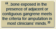
As a surgically trained Podiatrist, I am of the opinion that osteomyelitis should primarily be treated surgically. I have too many patients who have had long standing DFUs treated with numerous courses of antibiotics who still failed to heal their wounds due to persistent underlying osteomyelitis (yes, they were also properly debrided and offloaded and yes, they were treated with culture guided antimicrobial therapy). Only when the underlying bone (or joint) was resected did they go on to fully heal. I certainly believe that when a diabetic foot joint is probed or when joint fluid exudes from a wound (and cultures positive), it needs to be resected, because there is little chance of cure without surgery. Resection, however, does not always imply amputation. If a toe is not gangrenous or ulcerated beyond hope, we do try to avoid amputation by performing a simple metatarsal-phalangeal (MTP) joint resection. Occasionally, a superficial bone debridement followed by culture driven antibiotics will be sufficient to cure the bone infection. For calcaneal osteomyelitis, we will attempt a partial calcanectomy with adjunctive use of parental antibiotics. Although not with highest quality evidence, many surgeons also prefer to insert antibiotic-loaded beads made of cement or absorbable calcium sulfate. The latter have the advantage of being resorbable, although they tend to drain for some time afterwards. The antibiotic-loaded cement beads have the disadvantage of needing to be removed at some point. I am aware of no comparative trials that suggest the addition of antibiotic beads has superior outcomes to bone resection alone. Of course, bone exposed in the presence of adjacent or contiguous gangrene meets the criteria for amputation in most clinicians’ minds.
It’s Medical Too
But what of primarily medical management with prolonged antimicrobial therapy for the proverbial six weeks administered parenterally? Most of us were also taught that the latter was the accepted course of therapy. And which agent is best? Are there any specifically indicated agents? Is oral therapy efficacious if it is culture directed? Unfortunately, there is little to no high quality evidence available to answer these questions. There have been no randomized trials for osteomyelitis (especially in the diabetic foot) comparing oral to parenteral therapy or one agent versus another in this regard. No one agent has been proven superior to another for the management of this condition. Nonetheless, our European and Canadian Medical colleagues certainly believe that osteomyelitis in the diabetic foot can be effectively treated with antimicrobial therapy without the need for surgery (except perhaps for superficial bone debridement). The problem with most such reports is that the diagnosis of osteomyelitis is uncertain, lacking bone biopsies and/or cultures in many cases. This raises the question as to whether there could have only been soft tissue infection present in those successfully treated patients. Although I must admit with some incredulity that I find medical therapy alone somewhat attractive (especially for our very sick or ischemic patients), I am not yet convinced that it can be effective in most cases of diabetic foot osteomyelitis (as opposed to hematogenous osteomyelitis in children).
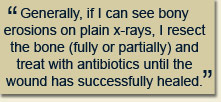
My current protocol for management falls somewhere in the middle of these two opposing schools of thought. As mentioned, when I see a metatarsal head and cartilage exposed at the base of a wound, I instinctively schedule it for a joint resection and culture guided antibiotics. While starting initially with intravenous therapy, I am now more comfortable sending the patient home on oral therapy, if appropriate agents for the pathogen are available. Calcaneal osteomyelitis I will routinely treat as mentioned earlier (with or without beads). Generally, if I can see bony erosions on plain x-rays, I resect the bone (fully or partially) and treat with antibiotics until the wound has successfully healed. As is often the case in the diabetic foot, bone is probed, x-rays are negative, but bone scans or MRI are positive. In such cases, I am now attempting to treat with good wound care and culture guided oral antibiotics (unless there might be a good reason for parenteral therapy). For hemodialysis patients with MRSA bone infection, I might administer parenteral vancomycin after each dialysis session.
Granted, my approach is largely anecdotal, but based on years of experience and many failures. Given the equipoise between the two opposing ways to treat osteomyelitis, I think a healthy combination of the two is probably the best approach to follow. I eagerly await prospective comparative studies or randomized trials to answer some of the questions posed and the conundrum that we routinely face in practice.
References are provided below that can expand upon many of the points made above. We welcome your opinions, concerns, and suggestions. If you have an interesting case or a troubling circumstance that you would like to share with fellow PRESENT Diabetes members, please feel free to comment on eTalk.
Best regards,

Robert Frykberg, DPM, MPH
PRESENT Editor,
Diabetic Limb Salvage
REFERENCES

-
Frykberg RG: An evidence-based approach to diabetic foot infections. American Journal of Surgery. 186 (Suppl 1):44-54, 2003
-
Frykberg RG, Wittmayer B, Zgonis T: Surgical Management of Diabetic Foot Infections and Osteomyelitis. Clinics Podiatr Med Surg. 24: 469-482, 2007
-
Thomas-Ramoutar C, Tierney E, Frykberg R : Osteomyelitis and Lower Extremity Amputations in the Diabetic Population. The Journal of Diabetic Foot Complications: 2010, 2 (1), No. 4, pp. 18-27.
-
Grayson et al: Probing to bone in infected pedal ulcers. JAMA March 1995 273: 721-723
-
Lozano et al: Validating the Probe-to-Bone Test and Other Tests for Diagnosing Chronic Osteomyelitis in the Diabetic Foot. Diabetes Care 33:2140–2145, 2010
-
Aragon-Sanchez J et al: Diagnosing diabetic foot osteomyelitis: is the combination of probe-to-bone test and plain radiography sufficient for high-risk inpatients? Diabet. Med. 28, 191–194 (2011)
-
Lew DP, Waldvogel FA. Osteomyelitis. Lancet364(9431):369-79, 2004.
-
Lavery L, Peters E et al: Risk factors for developing osteomyelitis in patients with diabetic foot wounds. Diabetes Research Clinical Practice 8 3 ( 2 0 0 9 ) 3 4 7– 3 5 2
-
Lipsky BA, Berendt AR, Deery G, et al: Diagnosis and Treatment of Diabetic Foot Infections. Clinical Infectious Diseases 2004;39:885–910
-
F. L. Game & W. J. Jeffcoate: Primarily non-surgical management of osteomyelitis of the foot in diabetes. Diabetologia (2008) 51:962–967
|
|