|
 Warren Joseph, DPM |
Negative Pressure wound therapy (NPWT) using the KCI Vacuum Assisted Closure (VAC) device is a well recognized modality to assist in the healing of complex wounds. The case presented in this article demonstrates its use in a complicated patient with osteomyelitis of the 2nd MTP joint who refused definitive cure by partial ray amputation.
The patient was a 51 y/o female, 231 lbs with insulin dependent diabetes mellitus and a long history of smoking and non-adherence with medical advice. She was admitted with the diagnosis of septic arthritis of the 2nd MTP joint with contiguous osteomyelitis of the base of the proximal phalanx of the 2nd toe and the head of the 2nd metatarsal. She had cellulitis of the forefoot with an open draining ulceration in the 1st interspace that probed to the joint.
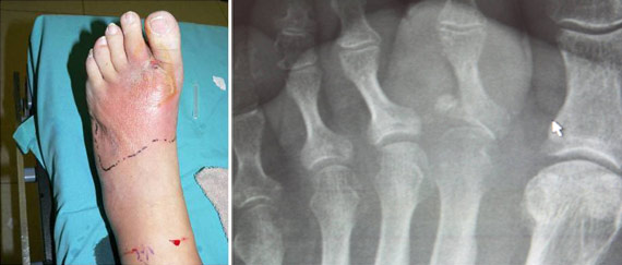
On the day of admission she was taken to the operating room and an incision and drainage was performed. The patient unequivocally refused partial ray amputation stating “If I wake up and my toe is missing I am going to sue the a** off the surgeon!” At the time of surgery minimal purulence was found. Bone around the joint was noted to be mildly necrotic. It was debrided and a sample was sent to pathology and microbiology. Vancomycin impregnated calcium sulfate beads were implanted.
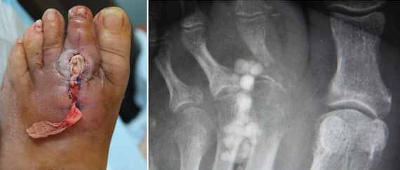
One day after the procedure a KCI VAC was placed onto the open surgical wound and over the implanted beads.
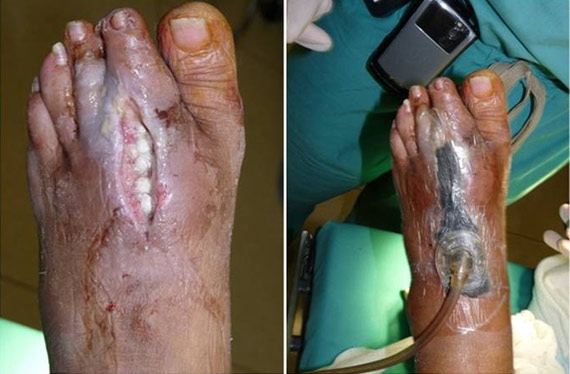
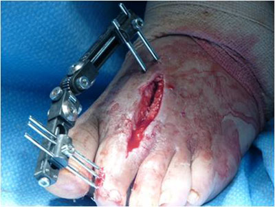
Cultures from bone and soft tissue grew a community associated MRSA (CA-MRSA) with a vancomycin MIC =1 and sensitive to both tetracycline and trimethoprim/sulfamethoxazole. Pathologic examination of the bone was read as being “consistent with osteomyelitis”. The patient continued to refuse partial ray amputation or, for that matter, even amputation of the toe. The VAC was continued as the wound was granulating well. The podiatric surgeon decided to return the patient to the OR for placement of an external fixator to maintain the alignment and length of the segment.
 |
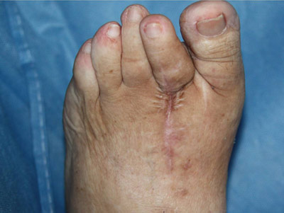
The patient was discharged from the hospital on oral doxycycline for 3 months. At 6 months the patient had totally healed the surgical site.
 |
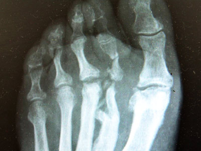
Although chronic bone changes were seen on x-ray they did not appear to be suggestive of an active osteomyelitis.
 |
At just over 1.5 years the patient has not had any evidence of infection and is clinically stable.
Although this case is not unique it does present some interesting clinical decision points:
-
The IDSA infection severity scale has become a standard used in most clinical trials and is being used in the clinical scenario to “get everybody on the same page” when it comes to the extent of infection and approach to treatment. In this case, the patient was graded as having an IDSA Moderate infection
-
How is osteomyelitis diagnosed? Although MRI may be the imaging standard, in this case plain film x-rays, pathologic examination of bone and, the true “gold standard”, positive bone culture were used without the need for more advanced imaging.
-
The patient refused “definitive” or ablative surgical treatment of her osteomyelitis. Recent evidence supports the use of more conservative surgery plus oral antibiotics as effective, as it was in this case.
-
After the initial I&D the patient needed dead space management. Certainly, the implantation of antibiotic beads may be useful, but to really get this wound to begin to close NPWT with the VAC was an important adjunct as it is in many of our diabetic foot infection patients following I&D and aggressive debridement.
|
###
|